信号, 软件和健康 01
医学成像 (Medical imaging)
- 定义: “医学成像指的是一个涉及专业仪器和技术的过程,用以创建关于身体内部生物结构和功能的图像或相关信息。” - A P Dhawan et al. (2008)
- 广义上,医学成像可以被认为是放射科学 (radiological science) 的一部分。
- 该领域本质上是多学科交叉 (multi-disciplinary) 的,需要来自多个学科的专业知识,包括:医学 (放射学)、物理学、化学、生理学、工程学和计算机科学。
解剖成像 vs 功能成像 (Anatomical vs Functional imaging)
医学成像可以根据其提供的信息类型分为两大类:
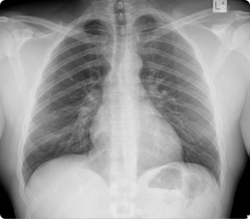
- 解剖成像 (Anatomical Imaging): 提供关于身体某一部位解剖结构 (anatomy or structure) 的信息。
- 例子: 胸部 X 光 (Chest X-ray),这可能是使用最广泛和最频繁的解剖图像模态。
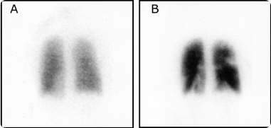
- 功能成像 (Functional Imaging): 检测或测量代谢 (metabolism)、血流、局部化学成分和吸收等方面的变化。
成像模态分类 (Anatomical vs Functional imaging)
| 解剖成像模态 (Anatomic imaging modalities) | 功能成像模态 (Functional imaging modalities) |
|---|---|
| X 射线 (X-ray) | 正电子发射断层扫描 (Positron Emission Tomography, PET) |
| 计算机断层扫描 (Computed Tomography, CT) | 单光子发射计算机断层扫描 (Single photon emission computed tomography, SPECT) |
| 磁共振成像 (Magnetic Resonance Imaging, MRI): 主要的 T1 和 T2 加权图像是解剖学的 | 光学荧光 (Optical fluorescence) |
| 超声 (Ultrasound) (多普勒除外) | 功能性磁共振成像 (Functional MRI, fMRI) |
| 光学成像 (Optical imaging): 包括光学相干断层扫描 (Optical coherence imaging, OCT) | 扩散张量成像 (Diffusion tensor MRI, DTI) |
| 其他 (Etc.) | 其他 (Etc.) |
界限的模糊:成像技术的融合
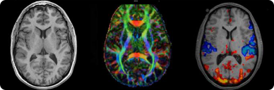
- MRI 与 功能性 MRI (MRI with T1 and functional MRI)
- 现代成像技术常常将解剖与功能信息结合起来。例如,MRI 可以提供多种信息。
图片说明:
- 左图: T1 加权 MRI (T1-weighted MRI),显示解剖结构。
- 中图: 扩散张量成像 (DTI),颜色表示纤维连接的主要方向。
- 右图: 功能性 MRI (fMRI),颜色表示血流中血氧水平的差异(暖色表示增加,冷色表示减少)。
- CT/PET
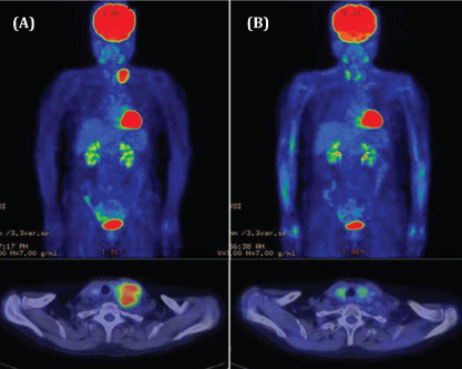
- CT/PET 融合是另一个典型例子,它将 CT 提供的精确解剖定位与 PET 提供的功能(代谢)信息相结合。
图片说明:
- (A) 一名患有肝细胞癌并转移至左锁骨上淋巴结的患者。
- (B) 同一位患者在针对该淋巴结进行放射治疗 3 个月后的情况,显示肿瘤活性显著降低。
医学成像的目的 (Purposes of medical imaging)
- 诊断 (Diagnosis)
- 研究 (Research)
- 术前手术规划 (Pre-operative surgical planning)
- 影像引导治疗 (Image-guided therapy)
- 诊断与治疗一体化 (Both diagnosis and therapy)
诊断 (Diagnosis)
获取特定器官或身体部位的医学图像以进行临床检查和诊断。
-
示例 1: 乳腺癌诊断 (Mammography)
- 通过乳腺 X 线摄影图像来诊断乳腺癌。
-
示例 2: 结肠息肉检测 (Colon polyps detection)
- 通过结肠 CT 进行结肠息肉检测(飞越式可视化)。
研究 (Research)
为研究目的提供信息,以研究正常及病理对象的解剖和功能结构。
-
示例 1: 阿尔茨海默病研究
- 通过脑部扫描对比阿尔茨海默病 (Alzheimer’s Disease) 患者与正常衰老 (Normal Aging) 个体的脑部功能差异。
-
示例 2: 以不同形式呈现数据
- 将成像数据以另一种形式呈现,例如将淀粉样蛋白沉积情况投影到大脑皮层表面进行分析。
说明: 诊断 (Diagnosis) 和 理解疾病机制 (understanding the disease mechanism) 是相互交织的过程。
术前手术规划 (Pre-operative surgical planning)
在手术前使用患者的影像数据进行模拟和规划,以确定最有效的手术方法。
-
示例 1: 神经外科手术模拟
- NeuroTouch Cranio: 模拟颅脑显微外科手术的系统。
- Dextroscope: 一种全息成像仪,将多模态患者图像融合成 3D 体积对象,可以进行分割和操作。
-
示例 2: 腹主动脉瘤的测量
- 通过 CT 图像进行腹主动脉瘤 (Abdominal Aortic Aneurysms, AAA) 的自动测量,用于支架植入规划。
影像引导治疗 (Image-guided therapy)
在治疗过程中实时使用医学影像来精确定位和引导治疗。
-
示例 1: 癌症的放射治疗 (Radiation therapy)
-
示例 2: 神经外科手术 (Neurosurgical operation)
- 使用增强现实 (Augmented Reality) 技术,将术中器械与患者的影像数据实时融合,实现精准导航。
诊断与治疗一体化 (Both diagnosis and therapy)
在某些介入性手术中,医学成像同时用于诊断和治疗。
- 示例: 心脏介入治疗 (Cardiac intervention)
- 通过血管造影术诊断冠状动脉的狭窄部位,并同时进行支架植入等治疗。
医学成像流程 (Medical Imaging process)
这是一个描述从成像原理到最终诊断或干预的完整流程的概念框图。
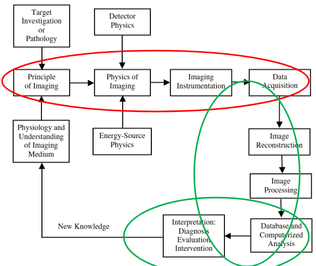
图片说明:
- Target Investigation or Pathology: 目标研究或病理学
- Detector Physics: 探测器物理学
- Principle of Imaging: 成像原理
- Physics of Imaging: 成像物理学
- Imaging Instrumentation: 成像仪器
- Data Acquisition: 数据采集
- Physiology and Understanding of Imaging Medium: 成像介质的生理学与理解
- Energy-Source Physics: 能源物理学
- Image Reconstruction: 图像重建
- Image Processing: 图像处理
- Interpretation: Diagnosis Evaluation Intervention: 解读:诊断、评估、干预
- Database and Computerized Analysis: 数据库与计算机化分析
- New Knowledge: 新知识
数字成像基础 (Digital imaging fundamental)
像素 (Pixel)
- 数字图像的最小元素。
- 图像大小通常以像素数量报告:
- MxN: 图像的高度 vs 宽度的像素数。
- 通常数字图像是光栅图像 (raster images):
- 像素网格是矩形网格。
- “在计算机图形学中,光栅图形图像是一种点阵数据结构 (dot matrix data structure),代表一个通常为矩形的像素网格,或颜色点,可通过显示器、纸张或其他显示介质查看。” (维基百科)
- 对于体积数据:单位是体素 (voxel)。
图像表示 (Image representation)
- 数字图像可以表示为矩阵 (matrices)。
- 每个像素都有一个值 (RGB 或 强度/灰度值 (intensity/gray value))。
强度图像 (Intensity image)
- 也称为灰度图像 (gray scale image)。
- 每个像素只有一个值 (ONE value) -> 图像是一个大小为 MxN 的矩阵。
- 通常,强度级别表示为整数:
- 类型 (Types):
uint8(0-255 强度级别),uint16, 等。
- 类型 (Types):
彩色图像 (Colour image)
- 红绿蓝 (Red Green Blue, RGB) 颜色格式 (非常常见) 或 色相饱和度值 (Hue Saturation Value, HSV) (有时有用)。
- 每个像素有一种颜色,由 R, G, B 的 3 个值编码 -> 每个像素有 3 个值。
- 图像实际上是一个三维矩阵 (3 dimensional matrix): MxNx3。
- RGB 或 HSV 值通常在 0 到 1 之间,但也可以是
uint8或uint16。
我们能用数字图像做什么?(What can we do with digital images)
- 增强 (Enhance): 提高图像对比度或清晰度 (例如:PET 图像超分辨率)。
- 滤波 (Filter): 去除噪声。
- 检测边缘/边界 (Detect edges/boundary)
- 分割 (Segmentation): 将图像划分为有意义的区域 (例如:脑肿瘤分割)。
- 压缩和重建 (Compress and reconstruction)
- 识别/分类 (Recognition/Classification): (例如:细胞图像分类)。
医学成像的元数据 - DICOM 格式 (Metadata for medical imaging- DICOM format)
- DICOM (Digital Imaging and Communications in Medicine,医学数字成像和通信) 是一个用于处理、存储、打印和传输医学成像信息的标准,由 NEMA (National Electrical Manufacturers Association,美国国家电气制造商协会) 开发。
- 包括文件格式 (file format) 和 通信协议 (communication protocol)。
- 通信协议定义了医学成像系统之间如何基于 TCP/IP 进行通信。
- 文件格式基本上有两个组成部分:
- 元数据 (Metadata)
- 图像数据 (Image data)
- 官网: http://dicom.nema.org/
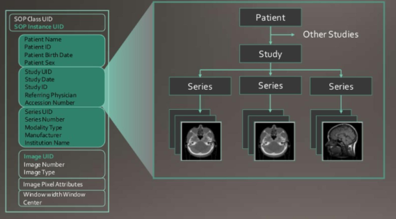
DICOM 元数据 (DICOM - Metadata)
DICOM 元数据可以包含大量信息:
-
图像模态类型:超声、MRI、CT、PET 等。
-
采集图像所用的协议:T1、T2、扩散张量等。
-
系统制造商和 ID:西门子 (Siemens)、飞利浦 (Philips) 等。
-
患者信息。
-
检查的日期、时间、持续时长。
-
图像数据格式:如
uint8、uint16等。 -
像素大小:如 3 mm x 3 mm。
-
切片厚度:如 5 mm。
-
序列中的切片/图像总数:如 512。
-
切片方向:轴位 (axial)、矢状位 (sagittal)、冠状位 (coronal) 等。
-
检查类型:如肺部检查。
-
以及更多信息。
-
元数据允许构建用于数字医学图像存储和检索的图像存档和通信系统 (Picture Archiving and Communication System, PACS)**。
-
同时,也实现了来自多个制造商的硬件之间的数据集成。
DICOM 数据结构
DICOM 图像 (DICOM - Image)
- 存储在 DICOM 文件中的图像通常具有以下格式之一:检查标签 (0002, 0010) (Transfer Syntax UID)
- JPEG
- JPEG lossless (无损)
- JPEG200
- 通常情况下,存储在 DICOM 文件中的图像是强度图像 (intensity image)。
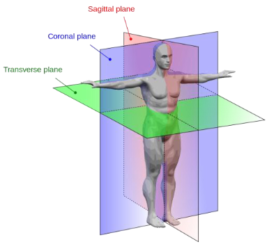
医学图像 - 解剖平面 (Medical image - anatomical plane)
- 医学图像的平面通常以解剖学术语 (anatomical terms) 来报告。
- 有 3 个(非常)常用的相互垂直的成像平面:
- 轴位平面 (Axial plane): 横断面 (transverse)
- 矢状位平面 (Sagittal plane): 中矢状面 (median plane), 旁矢状面平行于中矢状面但不在身体中线。
- 冠状位平面 (Coronal plane): 额状面 (frontal)
- 任何其他平面都是斜平面 (oblique plane),并且应指定该平面与一个垂直平面的夹角。
诊断性医学成像的效率测量 (Efficiency measures for diagnostic medical imaging)
- 医学成像最常用于辅助诊断决策。
- 医学成像模态的临床价值由以下指标确定:
| 真实情况 (Truth) | 合计 (Total) | ||
|---|---|---|---|
| 测试结果 (Test Result) | 患病 (Disease) | 未患病 (Non Disease) | |
| 阳性 (Positive) | A (真阳性, True Positive) |
B (假阳性, False Positive) |
$T_{Test \ Positive}$ |
| 阴性 (Negative) | C (假阴性, False Negative) |
D (真阴性, True Negative) |
$T_{Test \ Negative}$ |
| 合计 (Total) | $T_{Disease}$ | $T_{Non \ Disease}$ | Total |
- 敏感性 (Sensitivity): “敏感性是指在确实患有该疾病的人群中,测试结果为‘患病’的概率。” $$ Sensitivity = \frac{True \ Positive}{True \ Positive + False \ Negative} = \frac{A}{A+C} $$
- 特异性 (Specificity): “特异性是指在没有该疾病的人群中,测试结果为阴性的比例。” $$ Specificity = \frac{True \ Negative}{False \ Positive + True \ Negative} = \frac{D}{B+D} $$
- 阳性预测值 (Positive predictive value, PPV): 一个阳性测试结果者确实患有该疾病的概率是多少? $$ Positive \ predictive \ value = \frac{True \ Positive}{True \ Positive + False \ Positive} = \frac{A}{A+B} $$
- 阴性预测值 (Negative predictive value, NPV): 一个阴性测试结果者确实没有患该疾病的概率是多少? $$ Negative \ predictive \ value = \frac{True \ Negative}{True \ Negative + False \ Negative} = \frac{D}{D+C} $$
考点梳理与示例考题 (Review & Sample Questions)
本周内容作为导论,重点在于理解医学成像的基本概念、目的、分类以及相关技术标准。
可能考点:
- 医学成像的定义及其多学科性质。
- 解剖成像与功能成像的区别和联系,并能举例说明。
- 医学成像的五大主要目的(诊断、研究、术前规划、影像引导治疗、诊疗一体化)。
- 数字图像的基本概念(像素、体素、强度图像、彩色图像)。
- DICOM 标准的重要性、组成部分(元数据、图像数据)及其分层结构。
- 三个标准解剖平面(轴位、矢状位、冠状位)的定义。
- 诊断效率的四个核心指标(敏感性、特异性、PPV、NPV)的定义和计算公式。
示例考题 (Sample Questions)
问题 1: 请解释解剖成像 (Anatomical Imaging) 和功能成像 (Functional Imaging) 之间的主要区别,并为每种类型至少列出两种成像模态。
参考答案:
-
中文: 主要区别在于它们提供的信息类型。解剖成像提供关于身体部位物理结构和形态的信息,例如器官的大小、形状和位置。而功能成像则提供关于身体部位生理和代谢活动的信息,例如血流量、新陈代谢率或化学活性。
- 解剖成像模态示例: X 射线 (X-ray), 计算机断层扫描 (CT), 磁共振成像 (T1/T2 加权 MRI)。
- 功能成像模态示例: 正电子发射断层扫描 (PET), 功能性磁共振成像 (fMRI), 单光子发射计算机断层扫描 (SPECT)。
-
English: The main difference lies in the type of information they provide. Anatomical Imaging provides information about the physical structure and morphology of body parts, such as the size, shape, and location of organs. In contrast, Functional Imaging provides information about the physiological and metabolic activity of body parts, such as blood flow, metabolic rate, or chemical activity.
- Examples of Anatomical Imaging modalities: X-ray, Computed Tomography (CT), T1/T2-weighted Magnetic Resonance Imaging (MRI).
- Examples of Functional Imaging modalities: Positron Emission Tomography (PET), functional Magnetic Resonance Imaging (fMRI), Single Photon Emission Computed Tomography (SPECT).
问题 2: 什么是 DICOM?为什么它在现代医学中至关重要?
参考答案:
-
中文: DICOM 是“医学数字成像和通信 (Digital Imaging and Communications in Medicine)”的缩写。它是一套国际标准,用于定义医学图像及其相关信息的格式,以及在网络设备之间进行通信的协议。 它至关重要,原因有二:
- 互操作性 (Interoperability): DICOM 确保了来自不同制造商的医疗成像设备(如 CT、MRI、超声设备)和医疗信息系统(如 PACS、RIS)可以无缝地交换和使用数据。这避免了厂商锁定,促进了医疗机构内部及之间的协作。
- 数据完整性 (Data Integrity): DICOM 文件不仅包含图像数据,还捆绑了丰富的元数据(如患者信息、采集参数、检查详情等)。这确保了图像的上下文信息不会丢失,对于准确诊断、长期存档和临床研究至关重要。
-
English: DICOM stands for “Digital Imaging and Communications in Medicine.” It is an international standard for defining the format of medical images and their associated information, as well as the protocol for communicating them across networked devices. It is crucial in modern medicine for two main reasons:
- Interoperability: DICOM ensures that medical imaging equipment (like CT, MRI, ultrasound scanners) and medical information systems (like PACS, RIS) from different manufacturers can seamlessly exchange and use data. This prevents vendor lock-in and facilitates collaboration within and between healthcare institutions.
- Data Integrity: A DICOM file contains not only the image data but also a rich set of metadata (e.g., patient information, acquisition parameters, study details). This ensures that the contextual information of an image is not lost, which is critical for accurate diagnosis, long-term archiving, and clinical research.
问题 3: 假设一项新的医学影像测试被用于检测某种疾病。在 1000 名患者中,有 200 人确实患有该病。测试结果显示,180 名患病者测试为阳性,而 800 名未患病者中有 40 人测试为阳性。请计算该测试的敏感性 (Sensitivity)、特异性 (Specificity) 和阳性预测值 (PPV)。
参考答案:
-
中文: 首先,整理混淆矩阵:
- 总人数 = 1000
- 患病者 ($T_{Disease}$) = 200
- 未患病者 ($T_{Non \ Disease}$) = 800
- 真阳性 (A, True Positive) = 180
- 假阴性 (C, False Negative) = 200 - 180 = 20
- 假阳性 (B, False Positive) = 40
- 真阴性 (D, True Negative) = 800 - 40 = 760
计算:
- 敏感性 = $A / (A+C) = 180 / (180+20) = 180 / 200 = 0.9$ 或 90%。
- 特异性 = $D / (B+D) = 760 / (40+760) = 760 / 800 = 0.95$ 或 95%。
- 阳性预测值 (PPV) = $A / (A+B) = 180 / (180+40) = 180 / 220 \approx 0.818$ 或 81.8%。
-
English: First, organize the confusion matrix:
- Total patients = 1000
- With Disease ($T_{Disease}$) = 200
- Without Disease ($T_{Non \ Disease}$) = 800
- True Positives (A) = 180
- False Negatives (C) = 200 - 180 = 20
- False Positives (B) = 40
- True Negatives (D) = 800 - 40 = 760
Calculations:
- Sensitivity = $A / (A+C) = 180 / (180+20) = 180 / 200 = 0.9$ or 90%.
- Specificity = $D / (B+D) = 760 / (40+760) = 760 / 800 = 0.95$ or 95%.
- Positive Predictive Value (PPV) = $A / (A+B) = 180 / (180+40) = 180 / 220 \approx 0.818$ or 81.8%.